×
Single-use Bioprocess Products
Cell Culture Products
Protein Chromatography
Processing Filtration
Hydration Products
Single-Use Bioprocess Bags and Bottles
Single-Use Mixing System
Aseptic Connect & Disconnect
Bioprocess Supporting Vessels
Aseptic Sampling
Connectors and Fittings
Cell Culture Bioreactors
Cell Culture Bag
Microcarrier
Cell Sorting
Chromatography Column
Prepacked Column
Size Exclusion Chromatography Resin
Ion Exchange Chromatography Resin
Affinity Chromatography Resin
Hydrophobic Interaction Chromatography
Multimodal Chromatography
Polymer Chromatography
Filtration System
Filtration Filter
Filtration Holder
Water for Injection
Buffer Solution
Antifoaming Agent
BioHub® 2D Single-use Storage Bag
BioHub® 3D Single-use Storage Bag
NovaLinX® Nuclease-free Single-use Consumables
BioHub® Single-use Storage Bottle
BioHub® 3D Cubic Storage Bag
BioHub® 3D Circular Storage Bag
BioHub® Storage Bottle Tool Kit
BioHub® BM Single-use Magnetic Mixing System
BioHub® DS Single-use Split Desktop Mixing System
BioHub® UM Top-driven Mixing System
BioHub® FD Feeding System
BioHub® Single-use Filling System
BioHub® Fill Single-use Bulk Filling System
BioHub® Single-use Powder-feeding Bag
BioHub® MB Single-use Mixing Bag
BioHub® Single-use Weighing Bag
BioHub® Sealer Ⅱ
BioHub® Welder Ⅱ
BioHub® SC Sterile Cutter
Circular Plastic Bin
Plastic Freeze-thaw Box
Stainless Steel Freeze-thaw Box
Cubic Foldable Plastic Bin
Cubic Stainless Steel Tank
Storage Tray
Storage Tray Transfer Cart
BioHub® Single-use Sampling Bag
BioHub® Single-use Sampling Bottle
BioHub® S Sterile Sampling Base
BioHub® Connectors
BioHub® Sanitary Clamp Fittings
Biohub® Hose Plug
BioHub® Diaphragm Gasket
BioHub® Clamp & End Cap
BioHub® Integrated Diaphragm Gauge Tee
SynaLinX® B-Flex™ Thermoplastic Tubing
SynaLinX® TST Platinum Cured Silicone Tubing
CytoLinX® GB 1-20 L Benchtop Glass Bioreactor
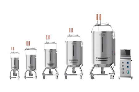
CytoLinX® BR 10-2000L Single-use Bioreactor
CytoLinX® WB Single-use Rocking Bioreactor
CytoLinX® WB Single-use Cell Therapy Bag
CytoLinX® WB Single-use Cell Culture Bag
CytoLinX® BRTF Single-use Top-driven Bioreactor Bag
Puredex® Cyto-1 Microcarrier
BioHub® Magnetic Separator
Chrom-LinX® Lab-scale Empty Chromatography Column
Chrom-LinX® Manual Compression Chromatography Column
Chrom-LinX® Automated Chromatography Column
PuriLinX® Benchtop Chromatography System
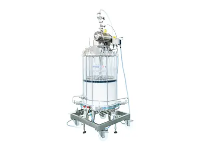
VERDOT Ips² InPlace™ Process-scale Chromatography Column
ChromaX® Geldex® Prepacked Column
Chrom-Trap® & Chrom-Screen™ Prepacked Column
ChromaX® A20 Affinity Chromatography Column
Chromstar® FF Resin for Size Exclusion Chromatography
Chromstar® CL Resin for Size Exclusion Chromatography
Chromstar® B Resin for Size Exclusion Chromatography
Geldex® PG Resin for Size Exclusion Chromatography
Puredex® G Resin for Size Exclusion Chromatography
Puredex® LH-20 Resin for Size Exclusion Chromatography
Maxtar® Resins for Ion Exchange Chromatography
Maxtar® HR Resins for Ion Exchange Chromatography
MaXtar® HR XL Resins for Ion Exchange Chromatography
Chromstar® BB Resins for Ion Exchange Chromatography
Chromstar® FF Resins for Ion Exchange Chromatography
Chromstar® HP Resins for Ion Exchange Chromatography
Chromstar® XL Resins for Ion Exchange Chromatography
Puredex® Resins for Ion Exchange Chromatography
MaXtar® ARPA Affinity Chromatography Resins
MaXtar® rProtein A Affinity Chromatography Resins
MaXtar® Protein G Affinity Chromatography Resins
rProtein A Chromstar® FF Affinity Chromatography Resins
Protein G Chromstar® 4FF Affinity Chromatography Resins
Protein G Chromstar® HP Affinity Chromatography Resins
Ni Chromstar® Affinity Chromatography Resins
IMAC Chromstar® Affinity Chromatography Resins
MaXtar® Chelating Affinity Chromatography Resins
Chelating Chromstar® FF Affinity Chromatography Resins
Glutathione Chromstar® Affinity Chromatography Resins
MaXtar® PlasmidCap HR Affinity Chromatography Resins
PlasmidCap Chromstar® HP Affinity Chromatography Resins
MaXtar® Heparin Affinity Chromatography Resins
Heparin Chromstar® Affinity Chromatography Resins
Benzamidine Chromstar® 4FF Affinity Chromatography Resins
MaXtar® Blue Affinity Chromatography Resins
Blue Chromstar® Affinity Chromatography Resins
NHS Activated Chromstar® 4FF Affinity Chromatography Resins
CNBr Activated Chromstar® Affinity Chromatography Resins
MaXtar® Hydrophobic Chromatography Resins
MaXtar® HR Hydrophobic Chromatography Resins
Chromstar® FF Hydrophobic Chromatography Resins
Chromstar® HP Hydrophobic Chromatography Resins
MaXtar® MMC Multimodal Chromatography Resins
Maxtar® COLL Multimodal Chromatography Resins
Hydroxyapatite BARONHAP® Type I&II Multimodal Chromatography Resin
Maxgo® 15S Polymer Chromatography
FiltraLinX® Lab TFF System
FiltraLinX® Pilot TFF System
FiltraLinX® Benchtop Manual Hollow Fiber TFF System
FiltraLinX® Benchtop TFF System
FiltraLinX® Automatic TFF System
FiltraLinX® Manual TFF System
FiltraLinX® Semi-automatic TFF System
FiltraLinX® Benchtop Manual Cassette TFF System
Cernolix® Series KSG Hydrophilic Filters
VentiLink® Series ESG Hydrophobic Filters
Puruflon® FSG Hydrophobic Filters
Cruclix™ Hollow Fiber Filter
FiltraLinX® Depth Filter Holder (Type P3C)
FiltraLinX® Depth Filter Holder (Type M)
FiltraLinX® Virus Filter Holder
FiltraLinX® Tangential Flow Filtration Holder
HydroLinX® Water for Injection in Bottles
HydroLinX® Water for Injection in Bags
HydroLinX® Hydration Products
HydroLinX® Antifoam Emulsion
HydroLinX® Buffer Solutions and Process Liquids
HydroLinX® Antifoam Emulsion

